


Varicose veins of the lower extremities are characterized by enlargement of the superficial veins of the legs, which is accompanied by a violation of the blood flow in it and a malfunction of the valves. As a result, the veins increase in length and diameter, having a zigzag, cylindrical or cuboidal shape, although there is also a mixed presentation of the listed malformations.
Features of the venous system
The emergence and development of varicose veins is directly related to the venous system of the legs, including:
- saphenous veins: small and large;
- deep veins (in the legs and thighs);
- perforated veins, which are the connecting link of the two previous systems.
Normally, 90% of blood is transported to the lower extremities through the deep veins, and the remaining 10% through the superficial veins. As it returns to one side of the heart, this mechanism is supported by valves in the walls of the veins. When the next blood arrives, they will slam shut to prevent its movement from top to bottom under the influence of gravity. The contracting muscles push more blood to the heart, allowing blood to flow normally.
When a person is in an upright position for a long time, blood can pool, increasing pressure in the veins and increasing their diameter. This process causes incomplete closure of the leaflets, resulting in disturbed blood flow with reverse flow from the heart - regurgitation.
The deep vein valves are most likely to be affected, as they transport the greatest amount of blood and therefore bear the maximum load. To reduce high blood pressure, part of the blood is transported by perforated veins to the superficial veins, which were not originally intended for a large volume. Such a load on the walls of the veins leads to their dilation and the formation of varicose veins.
At the same time, blood enters the deep veins without stopping, but due to a violation of their functions and the normal functioning of the valve leaflets of the perforated veins, blood is redistributed to the superficial vessels. face. As a result, chronic varicose veins develop, which over time is accompanied by painful sensations, edema and trophic ulcers.
Cause of disease
In the past, one of the main causes of varicose veins was called genetic predisposition, but today this theory has been disproved. Of course, it is possible to monitor frequent manifestations of the disease in some families, but this is more likely due to the peculiarities of life that are passed down in the family: culture of eating, passive rest, sedentary and the like.
The development of varicose veins is based on the presence of reflux in the venous system, when blood flows through the veins in the opposite direction. The transport of supplemental blood from deep veins to superficial veins may result from congenital or acquired degenerative pathology of the valve apparatus. This causes overfilling of the superficial vessels and their distension as venous nodes form.
One of the basic reasons for the development of varicose veins is considered an unhealthy diet, which in some cases leads to obesity. Such people move less, eat mostly highly processed foods, and the percentage of plant fiber in their diets is limited. After all, they are involved in strengthening the walls of veins and blood vessels and preventing prolonged chronic constipation, which significantly increases intra-abdominal pressure and thus causes varicose veins. It has been noted that an increase in body weight of more than 20% increases the risk of the disease fivefold.
The main trigger for women is being pregnant, while the risk of varicose veins increases with each subsequent pregnancy. Severe weight gain and growing uterus put stress on the legs, causing the body to stagnate. This condition is aggravated by the constantly increasing intra-abdominal pressure and the action of the hormone progesterone, which affects the state of elastic fibers in the walls of blood vessels.
Other factors that cause varicose veins of the lower extremities include:
- sedentary lifestyle, standing upright during the day (e. g. hairdresser), long flight or long journey. All this leads to stagnant processes in the lower extremities, when blood accumulates in the superficial veins and is poorly transported to the heart;
- sometimes increase the risk of varicose veins for women, wearing uncomfortable, tight shoes, especially high-heeled models;
- bras and underwear that are too tight will compress the veins in the groin area and increase the pressure in the abdomen, which is a direct premise leading to varicose veins;
- High Blood Pressure;
- Smoking, indirectly leads to thinning of the blood vessel walls.
Classification of diseases
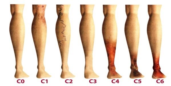
Varicose veins of the lower extremities are classified according to the prevalence of venous lesions, their localization, as well as the presence of pathological reflux, which is characterized by impaired blood flow. There are 4 types of varicose veins:
- Varicose veins in and under the skin (segment), in which no venous bleeding pathology;
- segmental varicose veins, when reflux occurs through perforated or superficial veins;
- a common form of varicose veins, in which reflux occurs through perforated and superficial veins at the same time;
- Varicose veins are characterized by reflux in the deep veins.
After varicose veins of the lower extremities become chronic, venology considers its three levels:
- Transient edema, occurring periodically against the background of the "heavy leg" syndrome.
- Persistent, prolonged edema. Hyperpigmentation and eczema may occur.
- Venous ulcers have a nutritional nature.
The latter is the most difficult to treat, as it requires preliminary removal of the inflammation and healing of the skin tissues.
Stages and symptoms
This disease develops very slowly, sometimes more than a decade has passed, until the symptoms appear, the patient is forced to seek medical attention. In the early stages of varicose veins, its manifestations are often attributed to fatigue, age, or other reasons. To fully consider the symptoms of the disease, its manifestations are classified according to the stages of varicose veins:
- The first stage begins to manifest itself more often at a young age - after the age of 20, when there is a feeling of heaviness in the legs, edema may appear and completely disappear overnight. On the inner side of the lower leg, you can see an enlarged vein, represented by a raised skin lump. At this stage, many people notice small spider veins. In general, the symptomatology is subtle and rarely receives the attention it deserves.
- The second stage is characterized by an increase in the external manifestation of varicose veins. The disease has developed against the background of pathological activity of the venous valves, as a result of which the hemispheric veins increase significantly in size, and their elongation can also be noted. Often presenting with heaviness and burning in the legs, they quickly get tired on long walks.
- This disease has become chronic due to the constant imbalance in the flow of venous blood. In the evening, the patient experiences swelling near the ankle, which can be very intense. Legs are heavy and cramps may occur at night.
- Without treatment in the earlier stages, the chronic functional deterioration of the venous system will negatively affect the metabolism of the skin, the areas of the lower legs are especially affected. Darkening of the skin visible near the ankles - hyperpigmentation, the skin thickens and becomes inflamed over time. The condition described is known as steatosis. If at this point you do not start treatment related to the venous system, then trophic ulcers will soon form.
- The fifth stage is accompanied by many trophic ulcers, some of them periodically healing with the formation of scars.
- In the area of long-lived nutritional disturbances, extensive ulcers open. This condition requires urgent active treatment, aimed at treating varicose veins and healing sores on the skin.
Diagnose
Perform an external examination of the lower extremities in longitudinal and transverse positions of the body, palpate the veins, and assess the preliminary stage of the disease. The patient is sent for a general blood test, which allows you to study the picture of the disease more thoroughly:
- at the platelet level, a tendency to thrombus formation will be reflected;
- the level of hemoglobin, as well as the number of red blood cells, indicates the degree of blood clotting;
- Because of the increased level of white blood cells, one can assess inflammation, helping to diagnose thrombophlebitis more quickly.
Be sure to examine the venous system of the legs, there are many methods:
- dopplerography ultrasound - USDG;
- phlebography;
- CT phlebography;
- dual circuit scan - USAS;
- phleboscintiography;
- clone photography;
- thrombus measurement and the like.
In practice, patients are more often prescribed USAS and USG, as they help to fully study the venous system of the leg and identify areas of degeneration. The remaining methods may be additionally indicated if the ultrasound examination does not provide a complete picture of the disease. Some of these methods may have complications such as venous thrombosis, catheter perforation, and allergy to the contrast medium. Consider the most commonly practiced techniques in phlebology:
- USAS allows the evaluation of anatomical, hemodynamic, and functional pathologies of the venous bed. The collected data will be processed by a computer, then the model of the venous system can be viewed on video or printed on paper.
- Doppler ultrasound with high accuracy determines the degree of intelligence of superficial and deep veins, blood flow velocity. Doppler ultrasound helps to evaluate the functioning of the valve apparatus.
After an extensive diagnosis, the doctor will take the patient's phlebocard, which will allow you to identify the damaged segments of the venous system, their extent and length. Then a suitable treatment is selected.
Treatment
It is done holistically and is determined based on symptoms, disease progression, and results of research. In the early stages, conservative therapy is prescribed, which includes:
- Drug treatment when a class of drugs is prescribed:
- anti-protective drugs and lung tonics;
- anticoagulants;
- separated person
- topical preparations (ointments, gels);
- anti-inflammatory drugs.
- Elastic compression, using knitted fabric or compression tape (rarely). It allows you to strengthen the contractions of the muscles, prevent stagnation, improve blood flow through the vessels. When wearing such underwear, there is an effect of artificially maintaining vascular tone.
- Physiotherapy methods, in which the best treatment results are shown by electrophoresis, electrodynamic currents, laser radiation and magnetic fields.
- Physical activity that is feasible, should only be done with compression underwear (except swimming). Cycling, swimming, jogging are recommended. The phlebologist chooses a separate group of exercises for the lower extremities, which will train the blood vessels of the legs every day.
Additionally, patients are advised to perform five-minute contrast procedures in the shower each evening, alternating from warm to cold water. Such manipulations improve blood flow and tighten blood vessels.
When initiating treatment, it is important to identify the causative factor in order to affect it effectively. And at-risk patients should visit a phlebologist every 2 years for a prophylactic examination and ultrasound of the leg veins.
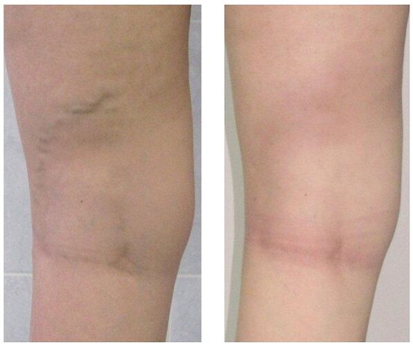
When conservative treatment is unsuccessful or varicose veins are observed at an advanced stage, surgical intervention is used. Today varicose veins can be completely cured by the following methods:
- Remove veins. The essence of the surgery is to remove the main trunks of the superficial veins to eliminate pathological bleeding. Perforated ribs are usually joined for the same purpose.
- Therapy therapy. It involves the introduction of sclerosus into the affected area of the vein, which leads to the connection of its walls. Recently, they began to actively use foaming agents for similar purposes according to technology -. Blood flow through the defect area is stopped and the cosmetic defect is eliminated in the form of a nodule. After such an intervention, leaving no scars, all manipulations were performed on an outpatient basis with no subsequent inpatient stay. But sclerotherapy is only used to fuse small branches of the venous line.
- Coagulation by laser. With the help of a laser beam, the marked vein is heated, the walls stick together and blood flow through it stops. But this technique is indicated only for veins with dilatation diameter less than one centimeter.
Prevent
Preventive measures can be primary, aimed at preventing the development of varicose veins, and secondary, when necessary, to reduce the risk of recurrence after surgery or to prevent disease progression. Helpful tips:
- lead an active lifestyle without putting heavy loads on your legs: swimming, walking, cycling;
- see your weight;
- keep both legs elevated more often;
- do not wear tight underwear and heels over 4 inches;
- use orthopedic insoles;
- contrast bath;
- do five-minute leg prevention exercises every day;
- Wear compression stockings for long walks.
If you notice the slightest suspicion of varicose veins - prominent nodules on your legs, swelling, worsening, do not postpone seeing a vascular doctor. Indeed, over time, this insidious disease can cause a host of complications, including thrombophlebitis and thrombophlebitis.